


Cosmetic Surgery in Dubai: The Ultimate Guide
Thinking about cosmetic surgery in Dubai? You’re not alone! Many people choose Dubai for their beauty transformations, but it’s natural to have questions: How much does it cost? Which clinic should I choose? Is Dubai really the best destination for cosmetic procedures?
The good news is that Dubai has established itself as one of the top destinations for cosmetic surgery. With internationally trained surgeons, world-class facilities, and cutting-edge technology, this city is an excellent choice for anyone looking to enhance their appearance.
In this ultimate guide, we’ll cover everything you need to know to make an informed decision. From understanding the cost and types of surgeries available, to finding the best clinics and recovery tips—we’ve got you covered.
Why Dubai? Dubai offers a unique blend of safety, luxury, and expertise, making it a top choice for those seeking cosmetic surgery. Whether you’re considering liposuction, tummy tuck, rhinoplasty, or any other procedure, Dubai has the professionals and resources to help you achieve the results you’ve always wanted.
Why Dubai is a Leading Destination for Cosmetic Surgery
If you’re considering cosmetic surgery, you might wonder, “Why Dubai?” Well, Dubai isn’t just famous for its stunning skyline and luxury shopping—it’s also become a global hub for top-tier medical services, especially when it comes to cosmetic surgery.
So, why should you choose Dubai for your beauty transformation? Here’s why Dubai stands out:
Experienced Surgeons: Dubai is home to internationally trained and board-certified surgeons who have years of experience performing cosmetic procedures with outstanding results.
Advanced Technology: With state-of-the-art clinics and the latest surgical techniques, Dubai provides access to world-class equipment, ensuring you get the best possible care.
World-Class Facilities: Dubai’s clinics offer luxurious, comfortable environments where you can relax and recover with the highest standard of care.
Safety and Standards: The city follows strict international medical regulations, so you can trust that your safety is a top priority throughout the entire process.
Accessibility and Convenience: Whether you’re coming from Europe, Asia, or the Middle East, Dubai is centrally located, making it easy to travel for medical procedures.
In short, Dubai combines cutting-edge medical technology with world-renowned expertise, making it a top choice for anyone considering cosmetic surgery.
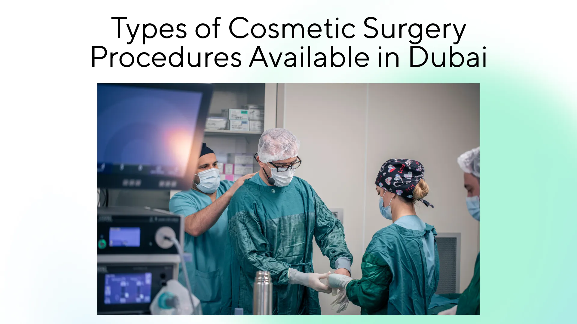
Types of Cosmetic Surgery Procedures Available in Dubai
Dubai offers a wide variety of cosmetic surgery options, catering to almost every aesthetic goal you might have. Whether you’re looking to enhance your body contour, rejuvenate your face, or improve your overall appearance, you’ll find a procedure that fits your needs. Let’s explore the most popular cosmetic surgeries performed in Dubai:
Liposuction in Dubai
Liposuction is one of the most requested procedures in Dubai. It’s perfect for removing stubborn fat from areas like the abdomen, thighs, or arms that are resistant to diet and exercise. The result? A smoother, more toned body shape.
Tummy Tuck Surgery in Dubai
If you’ve recently lost a lot of weight or had a baby, a tummy tuck (abdominoplasty) can help tighten loose skin and remove excess fat around the abdominal area. It’s a fantastic way to achieve a flatter, more sculpted stomach.
Rhinoplasty in Dubai
Rhinoplasty, or a nose job, is one of the most popular facial surgeries in Dubai. Whether you’re looking to refine the shape of your nose or fix a functional issue like a deviated septum, Dubai’s skilled surgeons can help you achieve your ideal look.
Breast Augmentation in Dubai
Breast augmentation is a procedure that enhances the size and shape of the breasts using implants. Whether you’re looking to increase volume or achieve a more balanced figure, Dubai offers world-class surgeons specializing in breast enhancements.
Facelift Surgery in Dubai
For those looking to rejuvenate their face, a facelift (rhytidectomy) helps smooth out wrinkles, tighten loose skin, and restore a youthful, refreshed appearance. It’s an excellent option for those looking to fight the signs of aging.
Dubai truly offers something for everyone when it comes to cosmetic surgery, and with top-notch facilities, you’re in great hands. Ready to take the next step? Let’s dive deeper into the costs and what to expect!
Cost of Cosmetic Surgery in Dubai: A Complete Breakdown
When planning for plastic surgery in Dubai, understanding the cost structure is key to making an informed decision. Dubai is renowned for offering high-end, world-class procedures, but costs can vary widely based on multiple factors. Let’s break it down clearly so you can plan ahead:
Overview of Cosmetic Surgery Costs in Dubai
Here’s a quick look at the average costs of some of the most popular cosmetic surgeries in Dubai:
| Procedure | Estimated Cost (USD) | Included Services |
|---|---|---|
| Liposuction | $3,000 – $7,000 | Surgeon fees, anesthesia, post-op garments, recovery support |
| Tummy Tuck | $5,000 – $10,000 | Surgery, anesthesia, hospital stay, post-operative care |
| Rhinoplasty (Nose Job) | $4,000 – $8,000 | Surgeon fees, anesthesia, follow-up visits |
| Breast Augmentation | $4,500 – $8,000 | Implant placement, anesthesia, follow-up care |
| Facelift (Rhytidectomy) | $7,000 – $12,000 | Surgery, anesthesia, post-op care, follow-up consultations |
What Affects the Price of Cosmetic Surgery in Dubai?
Here are key factors that influence the overall cost of your surgery in Dubai:
Procedure Complexity: More complex procedures like tummy tucks and facelifts naturally come at a higher cost due to the level of expertise required.
Surgeon’s Reputation and Experience: Highly sought-after, internationally trained surgeons might charge more, but their expertise often leads to better outcomes.
Clinic Location and Facilities: Clinics offering luxury services and premium facilities may charge more, but the quality and comfort of your experience are worth it.
Inclusion of Aftercare: Some clinics offer comprehensive post-surgery care in their packages, including follow-up consultations, recovery garments, and even luxury recovery suites.
Is Cosmetic Surgery in Dubai Worth the Investment?
While the costs may be higher than in some other countries, cosmetic surgery in Dubai is an investment in quality, safety, and long-lasting results. Here’s why:
World-Class Expertise: Dubai is home to some of the most skilled and experienced cosmetic surgeons globally.
Advanced Techniques and Equipment: Clinics use the latest technology and minimally invasive techniques, which often result in faster recovery and better outcomes.
Top-Notch Safety Standards: With strict regulations and international medical standards, Dubai ensures your safety and well-being throughout your procedure.
By understanding the cost breakdown and what’s included, you’ll be in a better position to make an informed decision about your cosmetic surgery in Dubai.
Dubai is emerging as one of the most trusted plastic surgery destinations. source
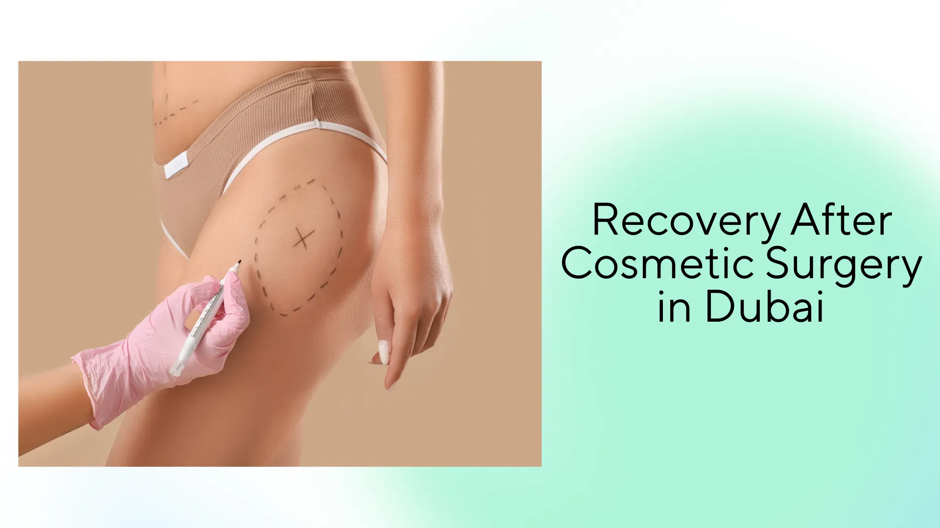
Recovery After Cosmetic Surgery in Dubai
Recovery is a crucial part of your cosmetic surgery journey in Dubai. Knowing what to expect during your recovery can make the whole process smoother and less stressful. Dubai’s clinics prioritize aftercare to ensure that you recover safely and quickly, so you can enjoy the results of your surgery with minimal downtime.
What to Expect After Cosmetic Surgery in Dubai
Recovery times vary depending on the type of surgery, but here’s a general overview:
| Time After Surgery | What to Expect | Key Recommendations |
|---|---|---|
| Days 1–3 | Swelling, bruising, and discomfort are common. You’ll need to rest and wear compression garments. | Take prescribed medications, avoid strenuous movements, and rest. |
| Week 1–2 | Light activities are possible, but some swelling and bruising may still be visible. | Follow post-op care, keep the incision area clean, and stay hydrated. |
| Week 3–6 | Significant improvement is visible. Most people can return to work or light exercise. | Continue wearing compression garments and avoid heavy lifting or strenuous activity. |
| 3–6 Months | Full recovery is reached and final results are visible. Any remaining swelling or bruising disappears. | Maintain a healthy lifestyle and attend follow-up consultations. |
Factors That Affect Recovery
Your recovery time can depend on several factors, including the type of surgery and your overall health. Here are some things to keep in mind:
Type of Surgery: Procedures like tummy tucks or facelifts usually require more time to heal compared to less invasive ones like liposuction. For patients seeking a more comprehensive transformation, a Body Lift in Dubai can be an excellent option, combining multiple procedures to achieve smoother, firmer contours with long-lasting results.
Surgeon’s Expertise: The more experienced your surgeon, the smoother your recovery is likely to be.
Health & Lifestyle: If you follow your surgeon’s post-op instructions, stay active within recommended limits, and eat a healthy diet, your recovery will be faster.
Following the surgeon’s aftercare plan is crucial to achieving the best results from your surgery. Make sure you attend all scheduled follow-up visits and take any prescribed medications as instructed.
Best Clinics for Cosmetic Surgery in Dubai
Choosing the right clinic for your cosmetic surgery in Dubai is key to achieving the best results. Dubai is home to many top-rated clinics offering world-class services and experienced surgeons.
Top Clinics for Cosmetic Surgery in Dubai
| Clinic Name | Specialty | Why Choose Them? |
|---|---|---|
| Aesthetica Clinic | Liposuction, Facelift | Expert surgeons, personalized care. |
| The American Academy of Cosmetic Surgery | Breast Augmentation, Tummy Tuck | Top-rated, luxurious recovery suites. |
| Cosmetic Surgery Dubai | Liposuction, Facelift | Affordable, high success rate. |
| European Aesthetic Surgery Clinic | Breast Augmentation, Rhinoplasty | Advanced technology and international standards. |
How to Choose the Right Clinic
Research Surgeon Experience: Ensure they specialize in your desired procedure.
Read Reviews: Patient feedback can give you insights into their satisfaction.
Consultation: Meet the surgeon to discuss your goals and expectations.
Do thorough research and choose a clinic with a proven track record to ensure the best outcome for your surgery.
Expertise of Cosmetic Surgeons in Dubai
Dubai is home to some of the best cosmetic surgeons, renowned for their expertise in procedures like liposuction, tummy tuck, rhinoplasty, and facelifts. These surgeons are internationally trained, bringing cutting-edge techniques and the latest technology to ensure natural-looking results with minimal recovery time. Whether you’re seeking body contouring or facial rejuvenation, Dubai offers world-class clinics and skilled professionals. To find top-rated surgeons, consider consulting the best cosmetic surgeons in Dubai who specialize in your desired procedure.
Start Your Journey with Expert Guidance
Cosmetic surgery in Dubai offers the perfect combination of advanced technology, skilled surgeons, and world-class clinics. Whether you are considering liposuction, tummy tuck, or even a full body lift, Dubai provides safe and reliable options with excellent results. To make the best choice for your personal journey, trust professionals who understand your needs. For expert guidance and access to top clinics, visit WMedTour and take the first step toward your transformation with confidence.
Frequently Asked Questions About Cosmetic Surgery in Dubai
1. How much does cosmetic surgery cost in Dubai?
The cost varies depending on the procedure. On average, surgeries like liposuction start from $3,000, while facelifts or tummy tucks can go up to $10,000 or more.
2. Is cosmetic surgery in Dubai safe?
Yes. Clinics in Dubai follow strict international safety standards, and most surgeons are board-certified with global training.
3. How long is the recovery period?
Recovery depends on the type of surgery. Less invasive procedures like liposuction take 1–2 weeks, while tummy tucks or facelifts may require up to 6 weeks.
4. Who is a good candidate for cosmetic surgery?
Healthy individuals with realistic expectations, stable weight, and no serious medical conditions are usually good candidates.
5. How do I choose the best surgeon in Dubai?
Look for board-certified surgeons, check patient reviews, and schedule a consultation to discuss your goals.