Description
What is Canaliculotomy?
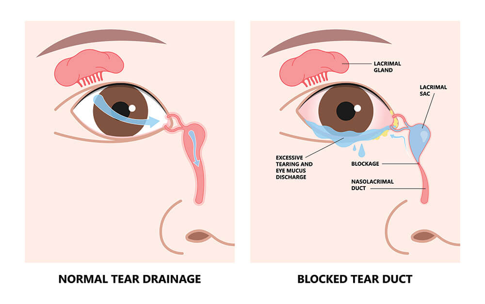
Canaliculotomy is a surgical procedure used to treat a blocked tear duct within the eyelid, specifically in the canaliculi – the tiny channels that drain tears from the inner corner of the eye (puncta) to the tear sac. Blockage in this area can lead to excessive tearing, recurrent infections, and discomfort.
Procedure:
Canaliculotomy is a minimally invasive outpatient procedure. Here’s a general breakdown:
- Anesthesia: Local anesthesia with numbing drops or a small injection is typically used to numb the area.
- Canaliculi Dilation: A very thin probe is inserted into the punctum to navigate the canaliculus. A balloon or other dilating tool may be used to open the blocked passage.
- Incision Creation: In some cases, a tiny incision is made using a laser or a blade on the inner lining of the lower eyelid, directly over the blocked canaliculus. This incision bypasses the blockage and allows tears to drain properly.
- Stenting (Optional): A small silicone tube (called a stent) may be inserted into the canaliculus to keep it open during healing.
Suitable Candidates:
Canaliculotomy is an option for individuals experiencing symptoms of a blocked canaliculus, such as:
- Excessive tearing (epiphora)
- Recurrent conjunctivitis (eye inflammation)
- Mucous discharge
- Localized discomfort or pain near the inner corner of the eyelid
- Failure of non-surgical methods like tear duct irrigation
Unsuitable Candidates:
Canaliculotomy may not be suitable for everyone. It’s generally not recommended for patients with:
- Uncontrolled active eye infections
- Severe scarring or damage to the canalicular system
- Unrealistic expectations about the outcome of surgery
- Blockage located deeper in the tear drainage system (beyond the canaliculi)
Advantages:
- Minimally Invasive: The procedure is relatively simple and quick, with minimal discomfort.
- Outpatient Procedure: Canaliculotomy is typically performed on an outpatient basis, allowing you to return home on the same day.
- Effective Tear Drainage: By addressing the blockage, the procedure helps restore proper tear drainage and alleviate associated symptoms.
Complications:
- Infection: Although uncommon, infection is a potential complication requiring prompt antibiotic treatment.
- Bleeding: Minor bleeding is possible but usually minimal.
- Punctal Stenosis: The tear drainage opening (punctum) can sometimes narrow again after surgery.
- Canalicular Injury: Accidental damage to the canaliculus during the procedure is a rare risk.
- Persistent Tearing: While less common, some patients may still experience occasional tearing after surgery.
Preoperative Care:
- Comprehensive eye exam to confirm the diagnosis and assess the tear drainage system.
- Dacryocystography (X-ray using contrast dye) or other imaging tests may be used to pinpoint the blockage location.
- Discussion of risks and benefits of canaliculotomy with your ophthalmologist.
- Stopping certain medications that could increase bleeding risk.
Postoperative Care:
- Eye drops or ointment to prevent infection and inflammation.
- Wearing an eye patch for a short period to protect the surgical site.
- Avoiding eye rubbing or straining for a few days.
- Stent removal (if used) at a follow-up appointment as directed by your ophthalmologist.
- Regular follow-up appointments with your ophthalmologist to monitor healing and address any complications.


Reviews
There are no reviews yet.