Description
Retinal Detachment Repair: Reattaching the Light-Sensitive Layer for Vision Restoration
Treatment Familiarity:
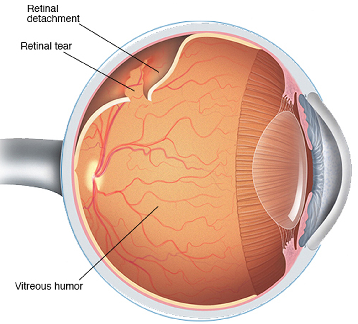
Retinal detachment repair is a surgical procedure performed by ophthalmologists (eye doctors) to reattach the retina, the light-sensitive layer at the back of the eye, when it pulls away from the underlying tissue. This detachment disrupts vision, and prompt treatment is crucial to prevent permanent vision loss. Retinal detachment repair is a relatively common procedure with high success rates when performed early.
Procedure Breakdown:
The specific approach for retinal detachment repair depends on the severity and location of the detachment. Here’s a general overview of some common techniques:
1. Pneumatic Retinopexy (Gas Bubble Placement):
- Anesthesia: Outpatient procedure using local anesthesia with numbing drops.
- Procedure: A small gas bubble is injected into the vitreous cavity (the gel-filled center of the eye). The bubble floats up and presses the detached retina back into place.
- Laser Treatment: Laser therapy may be used to create small burns around the retinal tear, creating scar tissue that helps hold the retina in place.
2. Scleral Buckle:
- Anesthesia: Local anesthesia with sedation or general anesthesia depending on the case.
- Procedure: A soft silicone band (buckle) is stitched onto the sclera (white part of the eye) to gently indent the eye wall and relieve pressure on the detached retina, allowing it to reattach.
- Laser Treatment: Similar to pneumatic retinopexy, laser therapy might be used to seal the retinal tear.
3. Vitrectomy (combined with other techniques):
- Anesthesia: General anesthesia is typically used.
- Procedure: Described previously (see Vitrectomy section), vitrectomy may be combined with other techniques like pneumatic retinopexy or laser treatment to address the detachment and any underlying issues within the vitreous cavity.
Suitable Candidates:
Retinal detachment repair is crucial for individuals experiencing symptoms of retinal detachment, such as:
- Sudden onset of flashes of light in one eye.
- A sudden increase in floaters (dark spots or squiggly lines) in one eye.
- A curtain-like shadow or loss of vision in one part of the field of vision (peripheral or central).
Who Might Not Be a Candidate?
While retinal detachment repair is essential for preserving vision, it might not be suitable for everyone in rare cases, such as:
- Individuals with very advanced detachment: If the detachment is very severe and has caused significant vision loss already, surgery may not be beneficial.
- People with severe medical conditions: Severe health problems that could increase surgical risk.
Advantages of Retinal Detachment Repair:
- Vision Preservation: Prompt surgical intervention can prevent permanent vision loss from retinal detachment.
- Improved Vision: Early successful repair can restore vision to near normal levels.
- Minimally Invasive Techniques: Advancements in surgical techniques have made some retinal detachment repairs less invasive with faster recovery times.
Potential Complications:
- Bleeding: Minor bleeding can occur during or after surgery.
- Infection: Although uncommon, infection is a potential complication requiring prompt antibiotic treatment.
- Cataract Formation: Retinal detachment repair can increase the risk of developing cataracts later in life.
- Increased Eye Pressure: Glaucoma, a condition of increased pressure within the eye, can develop after surgery.
- Re-detachment: There is a small risk of the retina detaching again, requiring additional surgery.
- Vision Changes: Temporary blurred vision or fluctuations in vision are common after surgery. Vision restoration may not be complete in all cases.
Preoperative Care:
- Comprehensive eye exam to assess the extent and location of the retinal detachment and overall eye health.
- Dilated eye exam for a detailed view of the retina.
- Imaging tests like ultrasound may be used in some cases.
- Discussion of risks and benefits of retinal detachment repair with your ophthalmologist.
- Medical evaluation to ensure you can undergo surgery safely.
Postoperative Care:
- Eye drops or ointment to prevent infection and inflammation.
- Wearing an eye patch or shield for a short period to protect the surgical site (depending on the procedure).
- Avoiding strenuous activity for a period of time, as instructed by your ophthalmologist.
- Regular follow-up appointments with your ophthalmologist are crucial to monitor healing, vision improvement, address any concerns, and detect potential complications early.


Reviews
There are no reviews yet.